photiomics: Focus on personalized cancer treatment
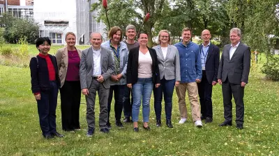
The consortium partners of the research project photiomics, which is funded by the German Federal Ministry of Education and Research, met in Hanover to present the results so far and agree on the next steps.

Bosch Research is working closely with industrial, academic, and clinical partners to advance personalized cancer treatment as part of the “photiomics” project, which is funded by the German Federal Ministry of Education and Research (grant number 13N15768). The established project consortium is developing an innovative diagnostic solution to test the effect of various cancer drugs on a patient’s tumor tissue and thereby determine a recommendation for personalized treatment. Since the project launch in July 2022, challenging research and development work has been successfully accomplished in the project with a volume of six million euros.
On September 25, 2024, a joint meeting of the project partners Bosch Research, Zellkraftwerk GmbH/Canopy Biosciences/Bruker Group (ZKW), Dr. Margarete Fischer-Bosch Institute of Clinical Pharmacology at Bosch Health Campus, Raylytic Software GmbH, and the project management VDI Technologiezentrum GmbH was held in Hanover.
At this joint meeting at ZKW, the consortium partners presented the current project status and the latest results of the ongoing research and development work. These include the cultivation system realized for tumor tissue slices, the immunofluorescence staining implemented, the 3D microscopy technology used, and AI-based image analysis methods. The technologies developed as part of the project and a successfully realized functional sample of a tumor bioreactor with a 3D microscopy system were presented in the laboratory. Together, the partners discussed the project findings and agreed on the next steps together.
The objective of the photiomics joint project
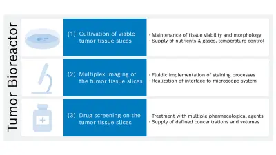
To increase the chances of success of drug therapy in cancer patients, the idea is to select drugs on a personalized basis before treatment begins. To do so, the effectiveness of various drugs is tested on a cancer patient’s living tumor tissue, which is available from a biopsy or surgery of the tumor. Thin tissue slices are then produced from the tissue, cultivated inside a tumor bioreactor, treated with drugs, and analyzed with a microscope.
The innovative and challenging objectives of photiomics are to develop a novel diagnostic laboratory system, the tumor bioreactor mentioned, and a method for conducting drug tests on tumor tissue sections using automated fluorescence imaging (4D: 3D and time) and AI-based analysis.
This promising approach makes it possible to study the reactions of living tumor tissue to different pharmacological treatments in an automated system. As a result, this can significantly improve the development of new drugs and therapies as well as research into diseases and relevant interactions, among other things.
The role of Bosch Research
In order to achieve these objectives, the Bosch Research strategic portfolio Healthcare Solutions is examining “cells-on-a-chip” technology within the scope of the project. An interdisciplinary team of scientists and engineers with complementary competencies in the fields of medical technology, physics, biology, chemistry, fluidics, and materials science is working on developing a system solution for an automated technology to cultivate tumor tissue in a controlled environment over several days. And with success: At the beginning of 2024, the achievement of this objective also marked the mid-term project milestone for the Bosch Research team.
In July 2024, the team transferred the cultivation system developed at Bosch Research and its associated components to the project partner ZKW in Hanover. The next project milestone was achieved there through collaboration. The cultivation system was successfully merged with the existing system for fluorescence microscopy in Hanover, which made it possible to document a living tissue slice completely and in three dimensions for the first time. Further research will now be carried out using the transferred system in Hanover.

Meanwhile, the team at Bosch Research in Renningen is working on further developing this system in order to realize the third function of the tumor bioreactor planned in the project: to conduct drug tests on tumor tissue slices using automated fluorescence imaging and image analysis with artificial intelligence.
Further information can be found in our Bosch Research Blog, in which research expert Dr. Bernd Scheufele explains the activities cells-on-chip technology and photiomics led by him in more detail.